How to maintain accurate bone geometries in your FEA meshes
by Dr. William Parr, University of New South Wales, Australia
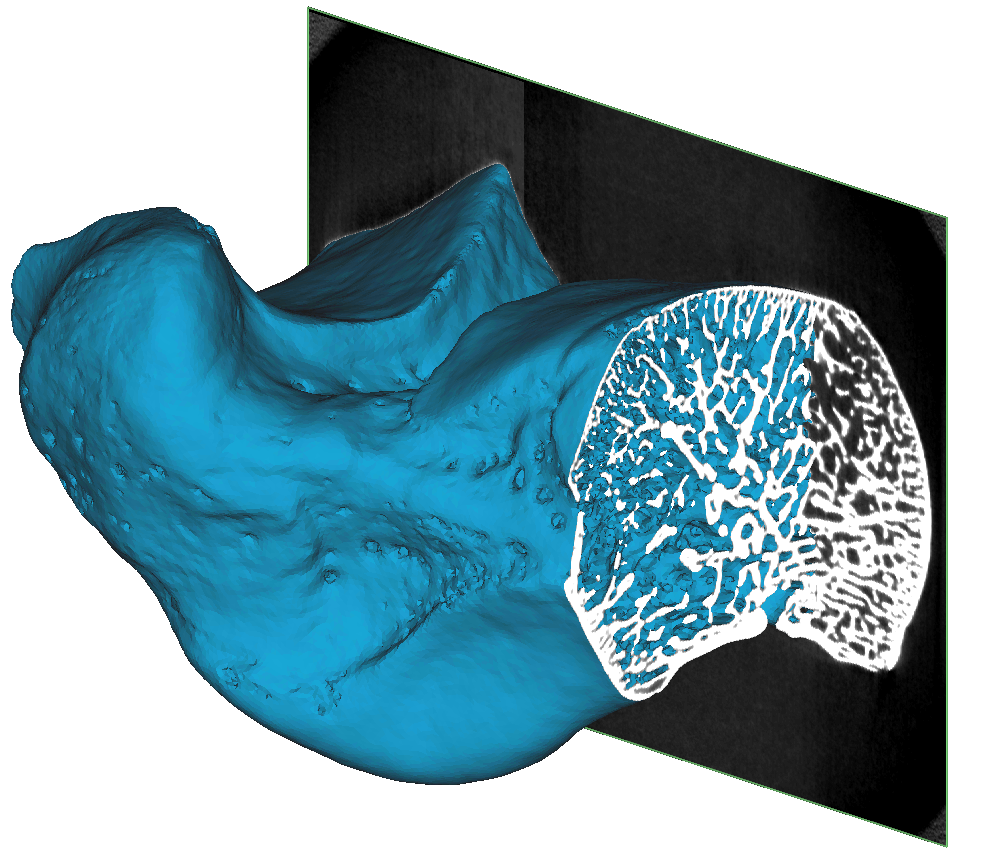
The ideal numerical solution is one that is mesh-independent, yet improved meshing algorithms would lead to more reliable FEA results. Bone consists of complex geometry with a cortical and trabecular structure. In most of the FEA analysis, the trabecular bone is represented as a bulky volume inside the cortical bone. However, since the trabecular bone has a complex inhomogeneous structure, this simplification could have a significant effect on the accuracy of your simulation.
Dr. William Parr wanted to compare these two approaches using a simplified trabecular bone geometry and the actual trabecular bone structure in the ankle. Due to the bone’s complex geometry, it is difficult to maintain geometrical accuracy while converting the micro-CT scan into a high-quality FEA mesh. This led Dr. Parr to collaborate with Materialise’s R&D team to find easier ways of creating complex trabecular bone FEA meshes.
During an engaging webinar, he provided an insight into his workflow and the results of his research.
Materialise medical device software may not be available in all markets because product availability is subject to the regulatory and/ or medical practices in individual markets. In countries where no regulatory registration is obtained of Mimics and/or 3-matic Medical, a research version is available. Please contact your Materialise representative if you have questions about the availability of Materialise medical device software in your area.
L-100786-01
This content is intended for Health Care Professionals only.