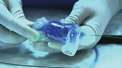
 Checking the balloon size in the 3D-printed model
Challenging Patient Anatomies
Though cardiac catheterization is becoming fairly common, the unique anatomy of each patient makes every procedure different from previous interventions. In addition, new devices are beginning to flood the market. With every new device, a patient puts his/her life in the hands of an interventionist, who is trying the procedure for the first time on a living patient. The three cases presented by Dr. AlJufan and his team are particularly challenging because the team was faced with very unique anatomies and hence the use of a new size of device. The ability to ensure fit and placement by simulating the procedure on an anatomically accurate 3D-printed model is particularly important for these types of procedures.
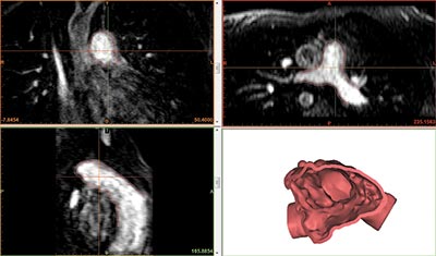 Screenshot of the Mimics Innovation Suite with the digital 3D model
Device Fit Can be Tested Again During the Procedure
Dr. AlJufan and his team implanted the 29 mm bioprosthetic valve from Edwards Lifesciences via cardiac catheterization. In all cases they were faced with a wide and challenging RVOT anatomy. The balloons were tested for effectiveness and fit in a 3D-printed model of each patient’s exact anatomy prior to and during the procedure to ensure an optimal device fit. Clinical engineers at Materialise used the Mimics Innovation Suite to produce the 3D-printed models of the patient’s complex RVOT.
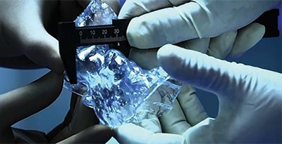 Checking the requirement measurements
-and-rigid-model-(right).jpg) Flexible model (left) and rigid model (right)
Flexible model (left) and rigid model (right)Meticulously Preparing a Complex Case
One procedure was extremely daunting because of the bulges and uneven characteristics of the RVOT. In addition, there was a very short landing zone. In the Mimics Innovation Suite, the patient’s anatomy was reconstructed from medical images so that the team could clearly see the complex RVOT. This digital model enabled them to analyze the ovality, deformities and presumed uneven tensile parts of the patient’s right ventricular track. Moreover, they could identify the landing zone of stenting and confirm the selected Percutaneous Pulmonary Valve Implantation (PPVI) size (29mm), the largest percutaneous implanted valve ever implanted. Once the model was 3D printed, Dr. AlJufan discussed the procedure with the patient, her family and his team.
Two models were offered by Materialise: a rigid model using the stereolithography technology as well as a flexible model using Materialise’s proprietary HeartPrint® Flex process. The team was able to take the required measurements on the rigid model and physically deploy the baloon in the flexible model. Once the baloon was deployed, Dr. AlJufan was even able to ‘tug’ on the balloon to be sure that the fit was tight. Besides, they also tested the stability of stenting, which is necessary for the 29 mm valve implantation.
“Not only were we able to perform a novel large-size valve implantation, but we could also cut down procedure time, shorten the recovery time and avoid an open heart surgery. Even though it was the first time we performed this kind of procedure, we felt confident. We now realize that this technology needs to be integrated in our cardiovascular treatment of structural heart disease.”
– Dr. Mansour AlJufan,King Faisal Specialist Hospital and Research Centre, Saudi Arabia
The Future of 3D Printing for Cath Labs
A growing number of cath labs is using 3D-printed models to prepare for their complex procedures. The beauty of 3D cardiac imaging and 3D Printing is that one can capture the anatomy at any point of the cardiac cycle and print both extremes from systole to diastole, as well as anywhere in between. Following Great Ormond Street Hospital and Guy’s and St. Thomas’ Hospital in London, King Faisal Specialist Hospital is one of the first centers to integrate this technology in treating structural heart disease outside the USA. Simulation of structural interventions helps in device fitting, improves accuracy and shortens the procedure time. It has the potential of personalized intracardiac device manufacturing.
 Dr. Mansour AlJufan and Dr. Fadel AlFadley
About Dr. Mansour AlJufan and KFSH&RC
Dr. AlJufan is a consultant cardiologist, Director of the Catheterization Laboratory at the Heart Center of King Faisal Specialist Hospital and Research Centre, Riyadh, Saudi Arabia. Trained at Boston Children’s Hospital and Massachusetts General Hospital in Boston, USA, he focuses on percutaneous repair of structural heart disease. He built his career with the support of Dr. Fadel AlFadley, Senior Cardiologist and Professor of cardiology, and still enjoys Dr. AlFadley’s mentorship. The KFSH&RC’s mission is to provide the highest level of specialized healthcare in an integrated educational and research setting.
Watch the live cases performed at PICS-AICS.