Why Point-of-Care 3D Printing?
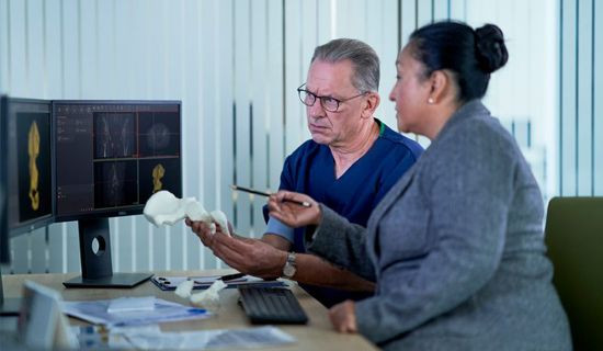
Go into the operating room with greater confidence
It has been proven that the risk of procedural complications can be reduced by planning procedures with 3D-printed models1. According to one study from the Mayo Clinic, 88% of oncologic surgeons find models ‘very likely’ to improve quality of care for patient2. Plus, by having an exact anatomical model, device selection and surgical equipment can better be determined in advance of the procedure.
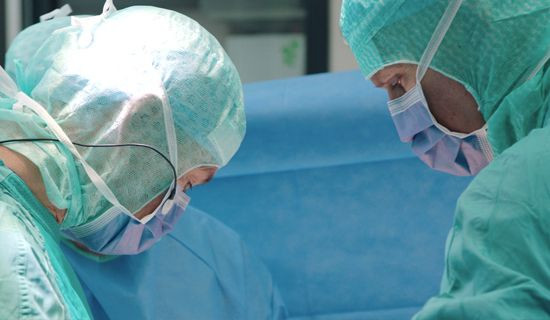
Reduce time and cost in the operating room
Procedure times can be shortened through the use of 3D-printed anatomical models to prepare for surgery3. Due to shorter surgery times, 3D-printed models have demonstrated a reduction in intraoperative radiation exposure by as much as 50%4. In addition, complication risk during surgery can be mitigated further reducing time in the operating room.

Increase patient safety, reduce length of stay and lower readmissions
Studies have shown that 3D-printed models can reduce the length of stay for patients receiving surgery for complex congenital heart procedures5.

Easily collaborate with multidisciplinary teams across the hospital
With Point-of-Care 3D Printing, you can enhance multidisciplinary team communication. This means that each person involved in a surgery – from radiologists to surgeons to supporting staff – can all understand the procedural approach.
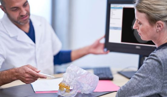
Achieve true informed consent
2D images on a computer screen can be difficult for patients to understand their anatomy and treatment options. A model in 3D that they can hold in their hand gives great insight to understand their own unique anatomy and buy-in to their treatment plan.
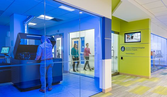
Stay abreast – and contribute – to cutting-edge care
Centralizing 3D printing in hospitals and bringing this capability to medical staff shows that you are at the forefront of medical innovation. With these tools, clinicians can then perform cutting-edge research and develop new therapies for future patients. In addition, 3D-printed models are a valuable training asset and can be used to teach the next generation of doctors in a more efficient and productive way.
1 Rogers-Vizena CR, Sporn SF, Daniels KM, Padwa BL, Weinstock P. Cost-Benefit Analysis of Three-Dimensional Craniofacial Models for Midfacial Distraction: A Pilot Study. The Cleft Palate-Craniofacial Journal. 2017;54(5):612-617. doi:10.1597/15-281.
2 Matsumoto JS, Morris JM, Rose PS. 3-Dimensional Printed Anatomic Models as Planning Aids in Complex Oncology Surgery. JAMA Oncology. 2016;2(9):1121. doi:10.1001/jamaoncol.2016.2469.
3 Rogers-Vizena CR, Sporn SF, Daniels KM, Padwa BL, Weinstock P. Cost-Benefit Analysis of Three-Dimensional Craniofacial Models for Midfacial Distraction: A Pilot Study. The Cleft Palate-Craniofacial Journal. 2017;54(5):612-617. doi:10.1597/15-281. Cherkasskiy L, Caffrey JP, Szewczyk AF, et al. Patient-specific 3D models aid planning for triplane proximal femoral osteotomy in slipped capital femoral epiphysis. Journal of Childrens Orthopaedics. 2017;11(2):147-153. doi:10.1302/1863-2548-11-170277. Zhao L, Zhou S, Fan T, Li B, Liang W, Dong H. Three-dimensional printing enhances preparation for repair of double outlet right ventricular surgery. Journal of Cardiac Surgery. 2018;33(1):24-27. doi:10.1111/jocs.13523.
4 Cherkasskiy L, Caffrey JP, Szewczyk AF, et al. Patient-specific 3D models aid planning for triplane proximal femoral osteotomy in slipped capital femoral epiphysis. Journal of Childrens Orthopaedics. 2017;11(2):147-153. doi:10.1302/1863-2548-11-170277.
5 Zhao L, Zhou S, Fan T, Li B, Liang W, Dong H. Three-dimensional printing enhances preparation for repair of double outlet right ventricular surgery. Journal of Cardiac Surgery. 2018;33(1):24-27. doi:10.1111/jocs.13523.
Resources
Contact us to find out how to have an end-to-end 3D printing at your Point-of-Care.